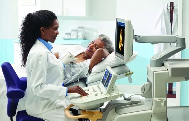
Sonography quietly saves lives every single day. This medical imaging modality plays a pivotal role in diagnosis, monitoring, and treatment planning across a range of medical specialties. In the world of sonography, every image tells a story, and every scan holds a key to better patient care. Whether tracking the progression of a disease or guiding a surgeon's hand with precision, medical sonographers are at the forefront of healthcare innovation.
Drawing insights from our conversation with Lacey Carpenter —MSRS, ARDMS (RDMS, RVT, RDCS), ARRT (R)(M)—who currently works with general, vascular, and obstetrics ultrasound, this guide delves into the diverse types of sonography and rewarding careers within this dynamic field, shedding light on its significance in modern healthcare.
Carpenter brings more than a decade of experience to her work as an ultrasound technologist, and her extensive credentials and wealth of expertise undoubtedly designate her as one-of-a-kind in the field. Beginning her career in rural Kansas, later becoming a traveling sonographer, and eventually settling in rural Colorado, she has humbly tended to underserved populations and communities with unwavering dedication over the years. Her versatility underscores her proficiency in this profession and ability to make a difference where it’s needed most.
What Is Sonography?
Sonography utilizes high-energy ultrasound waves to produce grayscale images from within a patient’s body, which are created by sending soundwaves into the body and capturing the echoes. This process delineates structures within the body, allowing for detailed examinations and helping diagnose and monitor various medical conditions.
Sonography vs. Ultrasound
The distinction between sonography and ultrasound, though often used interchangeably, lies in their application: Ultrasound is the technology, while sonography refers to the diagnostic process using this technology. In other words, an ultrasound is how the sonogram is produced. However, these medical professionals may refer to themselves as either medical sonographers or ultrasound technologists. Increasingly, ultrasound technologists and their schools are veering toward the title of “medical sonographers.”
How Does Ultrasound Work?
Ultrasound technologists use a transducer, which is similar to a camera, to capture images from soundwave reflections. It sends high-frequency soundwaves into the body and listens for sounds to bounce back. The machine creates the image based on how strongly they are returned. Most of these machines’ technology allows sonographers to adjust settings tailored to what part of the body is being examined.
For example, ultrasound waves interact with human tissue and are altered in multiple ways. The ways may change direction, slow down, speed up, or change frequency. In order to maximize image quality and correctly interpret the images, the sonographer must understand these interactions and adjust scanning parameters accordingly.
Along with the technical nuances of ultrasound equipment, this intricate process also requires a deep understanding of both internal and external human anatomy. Knowing what an image should look like surrounding specific body parts is crucial for identifying potential issues when something doesn’t look how it should. Oftentimes, ultrasound techs are already aware of a potential issue that doctors are suspicious of, like gallstones in the gallbladder or cirrhosis of the liver.
What about the gel substance used on patients’ skin during ultrasounds? Because sound does not travel as well through air, this gel serves as an acoustic coupling agent for sending the soundwaves directly into the body to prevent any air between the transducer and skin.
How to Become a Sonographer
The pathway to becoming a medical sonographer varies, from certificate programs to bachelor's and associate degrees to a combination of both. No matter which route you take, the importance of continuing experience and continuous education cannot be overstated when it comes to gaining specialized skills and certifications.
To be accepted into any of these programs, you will first need to apply. The American Registry for Diagnostic Medical Sonography (ARDMS) or Commission on Accreditation of Allied Health Education Program (CAAHEP) websites can help identify numerous accredited ultrasound schools in the United States that can equip you with the knowledge necessary to pass board exams. Once you pass that initial board exam, you may pursue additional certifications with continuing education.
If you didn't go through a specific ultrasound program but are already in the field, you could cross-train into other certain types of sonography by practicing your skills at work, then obtain your educational requirement through a continuing education course. From there, you must prove your clinical experience and skills in the area before you can qualify to take each test.
With that said, to become a certified ultrasound tech who is qualified to work involves a two-step process at minimum. First, you must take a physics board exam before pursuing more specialized credentials. This will essentially cover physics of the body, including blood flow and the physiology of anatomy. It’s also important to understand in depth how specific sonography machines and equipment work. Then, you must decide on at least one specialty to pursue and pass those relevant board exams.
Examples of specialization areas for medical sonographers include:
- Abdomen (AB)
- Breast (BR)
- Vascular technology (VT)
- Musculoskeletal (MSKS)
- Obstetrics and gynecology (OB/GYN)
- Fetal echocardiography (FE)
- Pediatric (PS)
- Adult echocardiography (AE) or pediatric echocardiography (PE)
- Midwife sonography
How Long Is Sonography School?
The journey to become a sonographer typically spans from two to four years, contingent upon the chosen educational path and desired level of expertise. A common trajectory with a two-year program might involve about a year of foundational coursework followed by a year of hands-on clinical experience. However, bachelor’s degrees extend the commitment to about four years; thus, two years could be a reasonable expectation for someone who simply wants to get their foot in the door.
Aside from the basic educational and credentials required to become an ultrasound tech, the importance of continuing education should not be understated. “You’re always going to be learning, and if you stop learning, you should probably stop your career,” Carpenter says. “You should always be looking at something new. With our degree, continuing education is required, so why not try to learn something new or gain more knowledge in those specific areas?”
Types of Ultrasound
The following types of sonography and specializations underscore its breadth and application across various medical fields.
Diagnostic Medical Sonography
Diagnostic medical sonography is an umbrella term covering the wide array of ultrasound specialties. This encompasses all forms of ultrasound, demonstrating the versatility of the field. Each sonographer, through their specialized training, contributes to the comprehensive diagnostic capabilities of medical teams. Those who earn a bachelor’s in this field may hold a degree title to the effect of “diagnostic medical sonography.”
Obstetric Ultrasound (Pregnancy)
For many, this specialty is often the first thing that comes to mind when they think of sonography. Obstetric (OB) ultrasounds are related solely to the pending birth of a baby. From confirming pregnancies to monitoring fetal development and well-being to identifying potential complications, obstetric ultrasound plays a central role in prenatal care from the first trimester through delivery. The personal and emotional excitement of these ultrasounds, coupled with their clinical importance, makes obstetric sonography a uniquely rewarding specialty.
Pelvic Ultrasound
Pelvic ultrasound complements obstetric sonography by assessing reproductive organs and aiding in diagnosing conditions related to infertility, pelvic pain, and irregular menstruation. Someone working in an OB clinic will typically have the skills to perform pelvic ultrasounds as well.
Musculoskeletal (MSK) Ultrasound
Musculoskeletal ultrasounds embody a bit more of an up-and-coming specialty focused on diagnosing conditions affecting muscles, joints, and tendons. The specificity required in imaging and interpreting musculoskeletal structures is increasingly valuable in sports medicine, rheumatology, and orthopedics. These professionals may work in orthopedic offices examining tears in rotator cuffs, tendons, ligaments, nerves, and the like. There is a required board exam for this specialization through the ARDMS.
Breast Ultrasound
This type of ultrasound is critical for evaluating breast lumps and pain, guiding biopsies, and assisting in the diagnosis of breast conditions, which could be anything from a normal cyst to breast cancer. Part of a breast ultrasound is the breast biopsy itself, and it’s common to use ultrasound instead of mammography for these biopsies because it’s easier for both the ultrasound tech and the patient.
Carpenter's expertise in both sonography and mammography underscores the collaborative approach to breast health and the role of ultrasound in real-time, guided biopsies. It is possible for mammographers to earn their credentials in breast ultrasound without completing a formal sonography program for a degree. They would need to do their clinicals and prove their skills, then take an educational course to help them pass the board exam.
View our Breast Ultrasound Course
Abdominal Ultrasound
Abdominal ultrasound is a cornerstone of medical imaging, allowing for the examination of vital organs such as the liver, kidneys, gallbladder, and spleen. This specialization requires a deep understanding of abdominal anatomy and pathology to identify conditions ranging from gallstones to liver disease. With a fundamental role in diagnostics surrounding the abdomen, this ultrasound is arguably the most common application within hospital settings.
Renal/Liver Ultrasound
This procedure in ultrasound allows for early detection and management of liver and renal conditions. Renal and liver imaging is essential for diagnosing diseases affecting these organs, such as cirrhosis and nonalcoholic fatty liver disease—which affects about 100 million people in the United States, per the American Liver Foundation. Liver scans can examine texture changes between a normal versus fatty liver as well as the blood flow into and out of the liver to get an idea of how it’s functioning. If a patient is experiencing liver failure and fluid collects in the abdominal cavity, ultrasound can be used to drain it.
Thyroid Ultrasound
Included within the abdominal imaging board exam, thyroid ultrasound entails evaluating thyroid nodules and guiding biopsies. The precision required in avoiding critical structures like the carotid artery during procedures exemplifies the sonographer's skill and the procedure's less invasive nature compared to traditional surgical methods. The patient is simply numbed around the area before the biopsy and leaves with only a bandage.
Vascular Ultrasound
Vascular ultrasound assesses the circulatory system and vessels in the body, identifying blockages and abnormalities in blood flow and ensuring proper circulation back to the heart. For instance, a sonographer can look at the vasculature in a patient’s stomach, arteries and veins in the leg, or carotid arteries in the neck. Having vascular credentials as an ultrasound tech is highly recommended and increasingly critical to comprehensive patient care.
View our Vascular Ultrasound Course
Cardiovascular Ultrasound
Cardiovascular ultrasound (AKA echocardiography) offers a detailed view of the heart's structure and function. It is essentially a live look at the heart. This ability to visualize heart muscle, contractions, and valve function in real time is crucial for cardiac care. Cardiovascular is a specialty a sonographer can pursue on its own, as many hospitals have dedicated echo departments.
Neurosonography
This type of sonography focuses on imaging the brain and nervous system, often using specialized windows through the skull to visualize cerebral structures and blood flow. This niche area of ultrasound requires precise knowledge of neuroanatomy and the challenges posed by the skull's bone density that protects the brain. There are specific spots where sonographers can look at the vessels in the circle of Willis, or the system of arteries providing blood flow to the brain.
Pediatric Ultrasound
Pediatric ultrasound is a specialized field tailored to meet the unique diagnostic needs of infants and children. It encompasses a broad range of applications, from assessing congenital anomalies to monitoring development and diagnosing conditions specific to pediatric patients.
Carpenter emphasizes the necessity of specialized training and certification for medical sonographers who wish to excel in this area, as there are distinct differences in imaging techniques and interpretations required when working with younger patients versus adults. This type of sonography calls for completing a specific pediatric ultrasound board exam, and these professionals often work in a children’s hospital or a larger facility with a pediatric unit.
Advance Your Career in Ultrasound With MTMI
Continuing education and certification in the dynamic field of sonography is not only essential to maintaining your skills; it is also required to advance and maintain your career. Ultrasound technologists must earn 30 credits every three years, although it does not need to be specific to any single specialty.
At MTMI, we offer multiple ultrasound training programs that help prepare sonographers and technologists for registry exams plus provide them with the knowledge to excel in their specialties. Choose a course and elevate your skill set as an ultrasound tech:
MTMI programs are taught by experts with national reputations in their fields and cover many modalities. Our cross-training courses offered in the classroom and via live webinars prepare you for registry exams and to take your career to the next level. Check out our full catalog of programs or contact us with questions today!


