- DocumentBreast Ultrasound Course Flyer for 2025 (296.98 KB)
Breast Ultrasound Course
About this Program
Sept. 26-28 Course in Denver has been changed to Webinar Only... MTMI students have a 97% pass rate when taking the board exam. There is no cure for cancer, but imaging teams can have an impact through screening.
Our breast sonography training course provides increased knowledge of the technical and clinical aspects of breast ultrasound and practical hands-on clinical experience in preparation for breast ultrasound certification. An overview of basic principles and techniques utilized in breast ultrasound is presented. This course is helpful for mammographers, sonographers, and radiologic technologists directly involved in breast sonography and imaging and physicians whose practice includes women’s health.
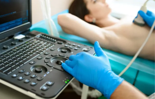
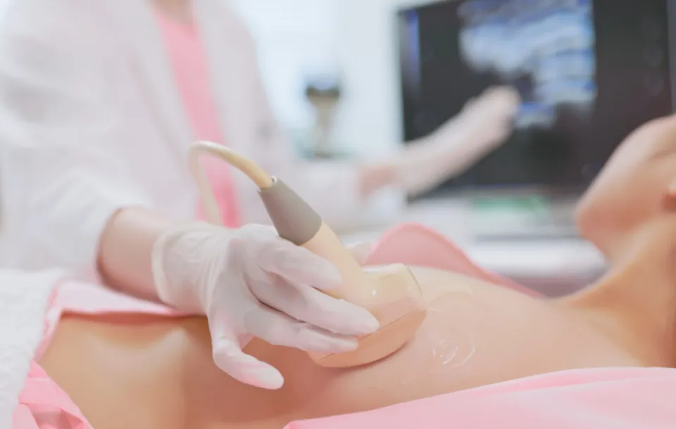
Our in-person and online breast sonography training courses utilizes a combination of lecture sessions and positioning workshops taught by an experienced breast sonographer and technologist. Instruction in patient scanning is stressed with hands-on demonstrations using clinical ultrasound scanners, tissue equivalent breast phantoms and live models. (Hands-on sessions are for attendees of in-person breast ultrasound classes only)
Plus, all attendees receive free breast ultrasound scanning videos to supplement learning.
This activity provides the 16 hours of structured education related to the content specifications outlined by the ARRT®, required for breast ultrasound certification and registration.
Educational Objectives
After this breast sonography training course, participants will be able to:
Optimize preparation for ARRT® and ARDMS Breast Ultrasound Registry exams through the SDMS
Fulfill breast ultrasound CME/CE requirements
Understand how the basic principles and instrumentation of ultrasound affect the technique of breast ultrasound
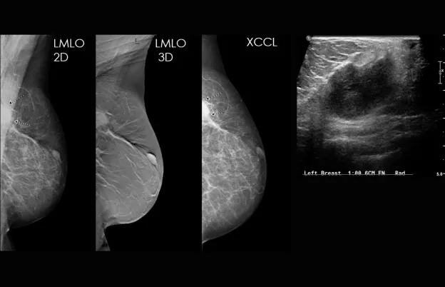
Recognize normal breast anatomy on the ultrasound image and evaluate lesions for their benign or malignant features
Recall the mammographic and ultrasonic features of specific benign and malignant lesions
Explain the role and importance of mammography, ultrasound, MRI, nuclear medicine, and ductography in the workup and management of the breast
Recognize breast ultrasound artifacts, understand why they occur, and describe techniques to minimize the occurrence of those that degrade the image
Develop an awareness of the invasive procedures that are available, as well as the benefits and limitations of each
Define the different surgical and nonsurgical treatment options for patients who develop breast cancer
Schedule
What this course will cover
Day 1
8:00 am – 8:30 am (Start times may vary based on location)
Introduction to Breast Ultrasound
8:30 am – 10:25 am
Patient Interactions and Management
- Accreditation of Ultrasound Facility and Personnel Certification Requirements
- Patient Communication
- Explanation of Procedure
- Patient Assessment
- Benefits and Limitations of Mammography and Ultrasound
- Introduction to Anatomy
- Safety Ergonomics
- Patient Positioning
- Image and Transducer Orientation
- Image Documentation
- Verification of Requested Examinations
- Determination of Appropriate Sequence
- Correlation of Imaging Request to Clinical Indications for Appropriateness
- Breast Cancer
- Epidemiology
- Signs and Symptoms
- Communication of Imaging to Supervising Physician
- Evaluation of Echo Patterns
- Review of Finding with Breast Cancer
10:25 am – 10:35 am | Break
10:35 am – 12:00 pm
Image Production
- Basic Principles of Ultrasound and Instrumentation Part 1
- Generation of Signal
- Ultrasound Wave Characteristics
12:00 pm – 12:45 pm | Lunch Break
12:45 pm – 1:20 pm
- Basic Principles of Ultrasound and Instrumentation Part 2
- Fundamentals
1:20 pm – 3:20 pm
Image Formation
- Selection and Adjustment of Technical Factors
- Safety and Bioeffects
- Other Imaging Tools
- Knobology and Technical Factors
3:20 pm – 3:30 pm | Break
3:30 pm – 5:00 pm
Scanning Lab 1
- Introduction to Breast Ultrasound
- Knobology
Basic/Foundations of Breast Ultrasound
Day 2
8:00 am – 9:55 am
Anatomy and Physiology
9:55 am – 10:05 am | Break
10:05 am – 11:30 am
Benign and Malignant Features
11:30 am – 12:00 pm
Evaluation and Selection of Representative Images
- Criteria of Diagnostic Quality
- Instrumentation Part 2
- Image Display & Storage
- Evaluation of Sonographic Equipment and Accessories
- Other techniques
- Artifact Recognition
- Instrumentation Part 2
12:00 pm – 12:30 pm | Lunch Break
12:30 pm – 1:00 pm
Evaluation and Selection of Representative Images (Continued)
1:00 pm – 2:30 pm
Procedures
- Pathology
- Benign Breast Lesions and Conditions
2:30 pm – 2:40 pm | Break
2:40 pm – 3:10 pm
- High-Risk Lesions and Conditions
- Atypical Ductal Hyperplasia
- Atypical Lobular Hyperplasia
- Papilloma w/Atypia
3:10 pm – 3:40 pm
- Quality Control
3:40 pm – 5:00 pm
Scanning Lab 2
- Positioning and Scanning Modification
- Nipple Techniques
- Axillary Scanning
Day 3
8:00 am – 9:00 am
Open Scanning (In-Person Attendees Only)
9:00 am – 10:00 am
Malignant Breast Lesions
- Lobular Carcinoma In-situ
- Ductal Carcinoma In-situ
- Invasive Ductal Carcinoma
- Paget’s Disease
- Invasive Lobular Carcinoma
- Other Uncommon Mammary Carcinomas
10:00 am – 10:10 am | Break
10:10 am – 11:10 am
Evaluation and Selection of Representative Images
- Criteria of Diagnostic Quality
- Demonstration of Anatomic Structure
- Demonstration of Pathologic Conditions
- Improvement of Sub-Optimal Images
11:10 am – 12:00 pm
Procedures
- Other Imaging Modalities
- BIRADs and Assessments
- DBT
- Ductography
- MRI
- CT & PET/CT
12:00 pm – 12:30 pm | Lunch Break
12:30 pm – 1:55 pm
- Other Imaging Modalities (continued)
- BSGI/PEM
- Augmented Breast Imaging
1:55 pm – 2:05 pm | Break
2:05 pm – 4:05 pm
Breast Interventions
- Image-Guided Procedures
- Cyst Aspiration
- Abscess Draining
- Fine Needle Aspiration
- Core Biopsy
- Vacuum-Assisted Biopsy
- Needle Localization/Surgical Biopsy
- Surgical Treatment and Changes
- Non-Surgical Treatment and Changes
4:05 pm – 4:35 pm
Emerging Technologies
- 3D Imaging
- Elastography
- Automated Breast Ultrasound (ABUS)
- Artificial Intelligence
Audience
Who should attend?
This course will be helpful for mammographers and sonographers who are directly involved in breast imaging and physicians whose practice includes women’s health.
Participants should understand the basic concepts of mammography and have an interest in breast ultrasound CME.
Program Faculty
Meet your presenter(s)

Sabah Aimadeddine
BS, RDMS
Sabah was born in Morocco and joined her husband in Texas in 1994. They have three grown daughters. Sabah majored in Microbiology and worked at Pfizer labs back in Morocco. After moving to USA, she wanted a job where she could interact and help people. Healthcare seemed the perfect place for her vision, so she went back to college for Radiographic Technology and later Ultrasound.
Sabah has been in the ultrasound field since 1999. She worked in many hospitals and has been with MD Anderson Cancer Center for the past 16 years; specializing in breast ultrasound. Her favorite activity is traveling.

Lacey Carpenter
MSRS, ARDMS (RDMS, RVT, RDCS), ARRT (R)(M)
Lacey Carpenter has more than a decade of expertise in breast imaging. She has been dedicated to serving the rural community in northeastern Colorado alongside her husband for nearly four years. They both have a deep affection for their community, where they are raising their daughter and cherishing quality time with family.
Before settling in Colorado, Lacey's career took her on a journey as a traveling sonographer across the Midwest of the United States, offering her expertise in General, Breast, Vascular, Obstetric, and Cardiac Ultrasound Imaging.
Lacey earned her Bachelor of Science in Medical Diagnostic Imaging with an emphasis in Radiology and Mammography in 2011. Continuing her commitment to excellence, while on her travels, she pursued further education and successfully obtained a Master's degree in Radiologic Sciences in 2019.

Michele Sisell
RT(R)(M)(BS)
Michele has been a registered radiologic technologist since 1998, specializing in mammography since 2005, and breast sonography since 2011. Michele worked at a community hospital in her hometown for over 22 years where she was the lead at the Women’s Health Pavilion breast imaging department. In 2020 she relocated to the Mayo Clinic in Rochester, MN in the breast Imaging and interventions department where she currently works. Michele enjoys sharing her knowledge which led her to be a clinical instructor in mammography and breast sonography for a local college. Michele’s passion for breast imaging and teaching led her to MTMI in 2018, where she became an independent breast imaging consultant helping facilities all over the United States with ACR failures, continuing education and specialized training in mammography and ultrasound. She enjoys teaching and mentoring others and feels blessed to be able to share her knowledge and experiences with others!
Credits
Accredited training programs
ASRT Category A
This program provides 23.5 hour(s) of Category A continuing education credit for radiologic technologists approved by ASRT and recognized by the ARRT® and various licensure states. Category A credit is also recognized for CE credit in Canada. You must attend the entire program to receive your certificate of completion.

CME
The Medical Technology Management Institute is accredited by the Accreditation Council for Continuing Medical Education to provide continuing medical education for physicians.
The Medical Technology Management Institute designates this live activity for a maximum of 23 AMA PRA Category 1 Credits ™. Physicians should claim only the credit commensurate with the extent of their participation in the activity
Location
Course location and hotels
Houston, TX - January 23 - 25, 2026
Accommodations & Course Location
Sheraton North Houston @ IAH
15700 John F. Kennedy Blvd
Houston, TX 77032
281.442.5100
Reservations: Click here
Rate: $132.00 + tax per night
Make reservation no later than:
January 08, 2026 to receive the discounted rate
Airport Transportation: IAH is 3.0 miles from the Sheraton.
Complimentary Hotel Airport Shuttle is available.
Reservation Required: 281.442.5100
Hotel Parking: Complimentary
Hotel Website: click here
| Milwaukee, WI- March 27-29, 2026 | |
Course Location MTMI International Headquarters | Accommodations: SpringHill Suites Milwaukee West Reservations click here Hotel Amenities: Airport Transportation: Springhill Suites does not provide airport |
| Wilmington, NC - April 24 - 26, 2026 |
Accommodations & Course Location Aloft Wilmington at Coastline Center Reservations: Click here Hotel Parking: $20.00 per day; Valet is available at $25.00 Hotel Website: click here |
| Milwaukee, WI- June 26-28, 2026 | |
Course Location MTMI International Headquarters | Accommodations: SpringHill Suites Milwaukee West Reservations click here Hotel Amenities: Airport Transportation: Springhill Suites does not provide airport |
| Milwaukee, WI- August 14-16, 2026 | |
Course Location MTMI International Headquarters | Accommodations: SpringHill Suites Milwaukee West Reservations click here Hotel Amenities: Airport Transportation: Springhill Suites does not provide airport |
| Las Vegas, NV- September 25-27, 2026 | |
Course Location/Accommodations: Reservations: Friday 09.25.26- Saturday 09.26.26 Rate Single/Double Occupancy Note: Booking through a 3rd party (such as Expedia, Priceline) will result in a $42.00 per night Hotel Parking: Complimentary Airport Transportation: Tuscany Suites hotel does not Hotel website click here |
Houston, TX - October 23 - 25, 2026
Accommodations & Course Location
Sheraton North Houston @ IAH
15700 John F. Kennedy Blvd
Houston, TX 77032
281.442.5100
Reservations: Click here
Rate: $132.00 + tax per night
Make reservation no later than:
October 08, 2026 to receive the discounted rate
Airport Transportation: IAH is 3.0 miles from the Sheraton.
Complimentary Hotel Airport Shuttle is available.
Reservation Required: 281.442.5100
Hotel Parking: Complimentary
Hotel Website: click here
Tuition

| Audience | Price | Early Price | Member Price | Member Early Price |
|---|---|---|---|---|
| Technologist | $899.00 | $859.00 | $869.00 | $829.00 |
| Physician | $1,179.00 | $1,129.00 | $1,149.00 | $1,099.00 |
| PA, APRN | $1,019.00 | $969.00 | $989.00 | $949.00 |
Early Pricing Guidelines
To qualify for Early registration rates, you must register at least 21 days before the program date.
Cancellation Policy
Courses
Refunds, minus a $50 processing fee, will be granted for cancellations received prior to 3 days before the program. Cancellations received within 3 days of the program will receive a credit toward a future MTMI program, minus the $50 processing fee. No refunds will be made after the program starts. MTMI reserves the right to cancel any scheduled program because of low advance registration or other reasons. MTMI’s liability is limited to a refund of any program tuition paid. MTMI recommends that attendees use refundable airline tickets. In case of cancellation of a program for any reason, MTMI is not responsible for travel costs incurred by attendees including non-refundable airline tickets. When offered, WEBINAR ATTENDEES that cannot log in due to unsolvable technical issues beyond their control will be eligible for a full refund.
