
Sub-Optimal Chest X-rays: Learn More, Do Better
About this Program
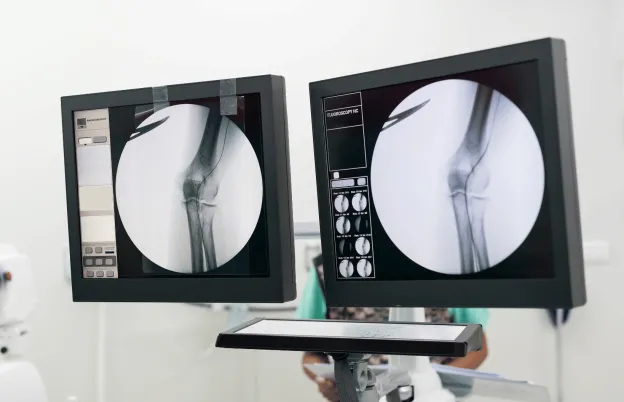
Chest radiography still represents the largest, single-most radiography examination ordered by practitioners even with all the choices available from the various medical imaging modalities. The simple chest radiograph delivers a high diagnostic yield of information, particularly when you consider its low cost and radiation dose, exam logistics and convenience.
In this session we’ll look at the basic interpretive techniques used by radiologists to understand the information seen on a chest image. Concepts such as Silhouette Sign, hilar overlay sign, cardiac curves, and chest consolidations will be discussed. The important role of the technologist will be emphasized to achieve optimum diagnostic yield
Educational Objectives
After this webinar, attendees will be able to:
- Explain the radiologic interpretive principle based on the five (5) radiographic densities
- Differentiate between the left and right hemi-diaphragm on a lateral chest radiograph, using the principle of five (5) radiographic densities
- Recognize abnormal chest radiographs using the silhouette sign concept
- Identify the general location of a chest consolidation using the silhouette sign and hilar overlay techniques
- Distinguish between the four (4) classes of radiographic chest consolidations
- Diagram the pulmonary acinus unit to explain the process of fluid collection and aeration in lung tissues
- Calculate the cardio-thoracic ratio as shown radiographically
- Label the left and right cardiac curves as seen radiographically
- Identify various interpretive anatomical structures commonly seen on a chest radiograph
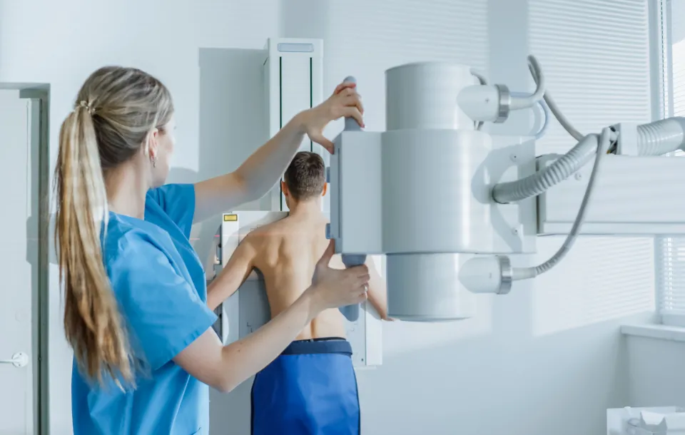
- Explain the important role the radiographer plays in optimizing chest image value and the consequences of sub-optimum image quality
Schedule
What this course will cover
- Tenets of Image Interpretation
- Five (5) Radiographic Densities and Image Production
- Silhouette Sign (SS)
- Principle of interpretation and history of SS
- SS and localization of pathologies
- Hilar overlay sign
- Spine sign
- Distinguishing between lt. and rt. Hemidiaphragms
- Continuous diaphragm sign
- Role of temporal resolution
- Classes of Chest Consolidations
- Tumor, fluid, atelectasis and pneumonia
- Benign vs. malignant tumors
- Chest fluids and infiltrates
- Congestive conditions and fluid
- Kerley lines, peri-bronchial cuffing
- Air-bronchogram and pneumonia
- Atelectasis (direct vs. indirect signs)
- Tumor, fluid, atelectasis and pneumonia
- Cardio-thoracic Ratio and Chest Analysis
- Calculation
- Cardiomegaly and typical causes
- Importance of patient positioning and inspiration
- Anterior clear space
- Retrocardiac clear space
- Heart Curves
- Left heart curves
- Right heart curves
- Heart curve accentuation and possible causes
- Chest Lines, Stripes and Anatomical Considerations
- Paratracheal stripes
- Hilar overlay sign
- Paraspinal line
- Chest Interpretation Strategies
- Alignment, rib interspacing
- Symmetry of aeration
- Consolidation
- Hyperlucency
- Cardiac silhouette
- Vascular markings
- Bronchial markings
- Masses and mass effect
- Volume loss
- Bony structures
- Role of Technologist in Optimizing Image Quality
- Importance of respiration
- Importance of positioning and rotation
- Image indicators of chest rotation
- Low-contrast resolution and exposure technique for chest radiography
Audience
Who should attend?
Radiologic Technologists
Program Faculty
Meet your presenter(s)

Randy Griswold
MPA, RT(R)
Randy has been in the medical imaging profession for over 40 years as an educator, sales and marketing professional and consultant. Currently he is a contributing author and medical imaging consultant. Prior to that, he was the Director of Sales and Marketing for a Midwest distributor of digital medical imaging products. His collective experiences as an educator include being the Program Director for Bellin College, School of Radiologic Sciences and their BSRS program. He was instrumental in transforming their two-year certificate program to a four-year, accredited BSRS degree program.
Randy has 23 years of experience in radiology capital equipment sales, service and support including digital imaging, and has completed his graduate work in Public Service Administration with an emphasis in Health Care Administration and Medical Imaging Marketing. He is a past president of the Wisconsin Association of Educators in Radiologic Technology (WAERT) and the Wisconsin Society of Radiologic Technologists (WSRT) as well as being a Fellow. His passion for teaching is focused on helping technologists understand the importance of obtaining good quality images for diagnosis, in a fashion that uses the skills and techniques of radiologist interpretation.
Credits
Accredited training programs
ASRT Category A
This program provides 2 hour(s) of Category A continuing education credit for radiologic technologists approved by ASRT and recognized by the ARRT® and various licensure states. Category A credit is also recognized for CE credit in Canada. You must attend the entire program to receive your certificate of completion.
Tuition

| Audience | Price | Early Price | Member Price | Member Early Price |
|---|---|---|---|---|
| Technologist | $37.50 | $33.75 | $32.50 | $29.25 |
Early Pricing Guidelines
Qualifying 'Early' registrations must be made at least 4 days in advance for the program.
Cancellation Policy
Webinars less than 8 hours of credit
Refunds, minus a $15 processing fee, will be granted for cancellations received at least 3 days prior to the program. Cancellations received within 3 days of the webinar will receive a credit toward a future MTMI program, minus the $15 processing fee. No refunds will be made after the webinar starts. MTMI reserves the right to cancel any scheduled program because of low advance registration or other reasons. MTMI’s liability is limited to a refund of any program tuition paid. WEBINAR ATTENDEES that cannot log in due to unsolvable technical issues beyond their control will be eligible for a full refund.