
Medical imaging or radiology is a rapidly growing field within the healthcare industry that offers a variety of exciting and rewarding career opportunities. From operating and maintaining high-tech equipment to interpreting images and working directly with patients, professionals in these fields play a crucial role in diagnosing and treating a wide range of medical conditions.
There are many different career paths available in medical imaging and radiology, each with its own unique set of responsibilities and requirements. Whether you're a recent high school graduate or a seasoned healthcare professional looking for a career change, there's never been a better time to explore the career options available in medical imaging and radiology. In this article, we'll take a closer look at some of the most popular career options for technologists in these fields, the skills and education required, and the job outlook for the future. To aid us in our exploration, we sat down with Jay Mazurowski, MS, CRA, FAHRA, and former President of Medical Technology Management Institute – MTMI Global.
What Is Medical Imaging?
Medical imaging refers to the use of various technological tools and techniques to produce visual representations of the internal structures of the body for medical diagnosis and treatment purposes. This can include a variety of diagnostic imaging modalities such as X-rays, computed tomography (CT), magnetic resonance imaging (MRI), ultrasound, and nuclear medicine scans. Each of these modalities produces different types of images that can be used to visualize various parts of the body, including bones, organs, and tissues, that can be used by medical professionals to identify and diagnose a wide range of medical conditions, from broken bones to cancer.
By analyzing these images, physicians can identify abnormalities or changes in the body that may be indicative of diseases, injuries, or other conditions. This information can be used to make accurate diagnoses and develop effective treatment plans for patients.
Medical Imaging Careers
Medical imaging careers offer a diverse range of opportunities for individuals interested in the healthcare industry. The field of medical imaging has experienced significant growth in recent years, and there is a high demand for skilled professionals who can operate sophisticated imaging equipment and work closely with patients and other healthcare professionals.
Radiographer / X-Ray Technologist
A radiographer, also known as an X-ray technologist, plays an essential role in diagnosing medical conditions by operating X-ray equipment, positioning patients, and capturing images of specific areas of the body. They work closely with physicians and other healthcare professionals to ensure accurate and precise imaging, as well as to communicate any findings or abnormalities that may be identified in the images. Radiographers must also ensure patient safety by following strict radiation protection protocols, such as wearing lead aprons and using collimators to minimize radiation exposure. Radiographers may also work with other imaging modalities, such as CT scans, MRI, and ultrasound, depending on their training, experience, and the requirements of their workplace.
According to Jay Mazurowski , “graduates of a two-year medical imaging and radiology associate degree program start as radiographers/x-ray technologists.” These early-career professionals can then go on to gain additional education and cross-training to pursue more advanced careers like the ones we’ll cover in the coming sections. Radiographers may also perform these more specific types of radiography, such as:
Fluoroscopy
Fluoroscopy is a type of medical imaging that uses a continuous X-ray beam to produce real-time moving images of the internal structures of the body and is often used during medical procedures such as surgery, cardiac catheterization, or gastrointestinal studies. The images produced by fluoroscopy can help healthcare professionals to guide instruments, such as catheters or needles, to the correct location within the body or monitor the progress of a procedure in real time.
Bone Densitometry
Also known as dual-energy X-ray absorptiometry (DEXA or DXA), bone-density X-ray is a type of medical imaging that measures the density of bones to diagnose and monitor conditions such as osteoporosis. During a bone density X-ray, the patient lies on a table while a scanner passes over the body, emitting low-dose X-rays that are absorbed differently by bone and soft tissue. The amount of X-ray energy that is absorbed by the bone is measured and used to determine the density of the bone.
To become a radiographer, individuals typically need to complete an accredited educational program and pass a certification exam.
MRI Technologist
An MRI technologist specializes in operating MRI equipment to produce images of the body's internal structures. MRI is a medical imaging modality that uses a strong magnetic field, radio waves, and a computer to produce detailed images of the body's organs, tissues, and bones. MRI technologists play a crucial role in ensuring that the MRI scanner is properly calibrated, positioned, and operated to produce accurate and high-quality images. They work closely with radiologists and other healthcare professionals who interpret the images and diagnose medical conditions. In addition to operating the MRI equipment, MRI technologists also ensure patient safety by screening patients for contraindications, such as pacemakers or metallic implants. They also ensure that patients are positioned safely and comfortably during the scan.
MRI technologists typically have a degree or certificate in radiologic technology or MRI technology and may also be certified in MRI, which requires MRI training credits. They may also need to meet licensing requirements for their state. MRI techs work in various settings, including hospitals, clinics, and imaging centers.
Read our blog all about becoming an MRI Technologist.
CT Technologist
A CT technologist is a healthcare professional specializing in operating Computed Tomography (CT) scanners. CT scans use X-ray and computer technology to produce detailed cross-sections of the body, which offer more detailed views of internal structures like bones, organs, and soft tissues. CT technologists are crucial to keeping patients safe while providing accurate imaging to be used by physicians in the diagnosis and treatment of a variety of medical conditions.
Completing required clinical training hours and being certified by the American Registry of Radiologic Technologists (ARRT®) is also required. Additionally, certain states may require licensing to practice as a CT technologist.
Read our blog all about becoming a CTTechnologist.
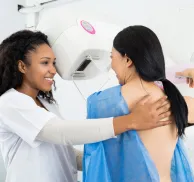
Mammographer
A mammographer specializes in producing medical images of the breasts using mammography, a specific type of X-ray imaging, which is used to detect and diagnose breast cancer and other breast abnormalities. Mammographers operate specialized mammography equipment that produces low-dose X-rays to capture images of breast tissue. They carefully position both the patient and the mammography equipment to acquire the required images. This helps ensure that the resulting images are of high quality and provide clear information for the radiologist to make an accurate diagnosis.
To become a mammographer, individuals must complete a certificate or associate degree program in radiologic technology and meet all of the MQSA requirements, including initial mammography training. They may work in a variety of settings, including hospitals, imaging centers, or clinics that specialize in breast health.
Read our blog all about becoming a Mammography Technologist.
Diagnostic Medical Sonographer
Diagnostic medical sonographers, also known as ultrasound technologists or sonographers, specialize in producing medical images using ultrasound technology. Ultrasound is a non-invasive, radiation-free medical imaging modality that uses high-frequency sound waves to create images of the body's internal structures. Sonographers operate specialized ultrasound equipment that emits sound waves and captures the returning echoes to produce images of organs, tissues, and blood flow within the body. They position the patient and the ultrasound equipment to obtain the necessary images and work closely with physicians and other healthcare professionals who interpret those images and diagnose medical conditions.
To become a diagnostic medical sonographer, individuals typically complete an associate or bachelor's degree program in diagnostic medical sonography and must also become certified. They work in various settings, including hospitals, clinics, imaging centers, and physician offices.
Diagnostic medical sonographers commonly perform abdominal ultrasounds but may specialize in specific areas, including:
Breast Ultrasound
Breast ultrasound is a medical imaging modality that uses high-frequency sound waves to produce images of the internal structures of the breast. Breast ultrasound is often used as a complementary imaging modality to mammography for breast cancer screening, diagnosis, and follow-up. Unlike mammography, breast ultrasound does not use radiation and is considered safe for pregnant and breastfeeding women. Breast ultrasound training is available for sonographers interested in becoming certified and registered in this specialty.
OB/GYN
OB/GYN ultrasound produces images of the female reproductive system, including the uterus, ovaries, and fetus, during pregnancy. OB/GYN ultrasound can be used to diagnose and monitor a wide range of medical conditions, including ectopic pregnancy, uterine fibroids, polycystic ovary syndrome (PCOS), and gynecologic cancers, and is an essential tool for obstetricians, gynecologists, and other healthcare professionals who specialize in women's health.
Pediatric
When sonography is used to examine the organs and tissues of children and infants, it’s called pediatric sonography. Pediatric sonography is used by pediatricians, neonatologists, and other healthcare professionals who specialize in caring for infants and children to diagnose and monitor a wide range of medical conditions, including congenital heart defects, kidney disease, liver disease, and abdominal tumors.
MSK (Musculoskeletal)
MSK, or musculoskeletal, ultrasound produces images of the body's muscles, tendons, ligaments, joints, and bones. This non-invasive imaging technique can be used to evaluate the musculoskeletal system and diagnose conditions, guide certain procedures, and monitor the progress of treatments. It is an essential tool for sports medicine physicians, orthopedists, rheumatologists, and those who specialize in the diagnosis and treatment of musculoskeletal conditions. MSK ultrasound courses are available to provide hands-on scanning experience to both physicians and technologists with varying amounts of MSK experience .
For more information on MSK, read our Comprehensive Guide to Musculoskeletal Ultrasound.
Vascular Sonographer
Vascular ultrasound does use ultrasound technology but is often separated from conventional ultrasound and diagnostic medical sonographers due to unique certification & registration. It can be used to evaluate the structure and function of the body's arteries and veins, including those in the neck, arms, legs, and abdomen, to diagnose and monitor medical conditions such as peripheral arterial disease, deep vein thrombosis, carotid artery disease, and abdominal aortic aneurysm. It can also be used to evaluate blood flow to organs and tissues and to guide certain procedures, such as the placement of catheters or the insertion of vascular access devices. An approved vascular ultrasound course will help you meet the structured education requirements for ARRT® certification and registration.
Nuclear Medicine Technologist
A nuclear medicine technologist is a healthcare professional who specializes in using radioactive materials, called radiopharmaceuticals, to produce images of the body's internal structures and functions, such as the heart, lungs, liver, and bones. Nuclear medicine technologists prepare and administer radiopharmaceuticals to patients, operate specialized imaging equipment, and assist physicians in making accurate diagnoses. They also monitor patients for adverse reactions to the radiopharmaceuticals and follow strict radiation safety protocols to minimize radiation exposure to patients, staff, and the public. Nuclear medicine technologists work in a variety of settings, including hospitals, clinics, and imaging centers. To become a nuclear medicine technologist, individuals typically complete an accredited educational program and pass a certification exam. They may also need to obtain a state license to practice nuclear medicine technology.
Alternative Jobs for Radiologic Technologists
In addition to the radiologist careers listed above, there are also alternative jobs available in the field of medical imaging and radiology. These jobs may require additional education and training, but they can offer new opportunities for career growth and advancement. Some examples include:
Imaging Informatics
Imaging informatics professionals are responsible for managing and analyzing the vast amounts of data generated by medical imaging technologies. They use specialized software and tools to organize, store, and analyze medical images and help healthcare professionals make more informed decisions.
PACS Administrator
Picture archiving and communication system (PACS) administrators are responsible for managing and maintaining the computer systems used to store and transmit medical images. They make sure the systems are functioning correctly and work with healthcare professionals to ensure that images are accessible when needed. PACS admin courses are available to provide the essential skills for the implementation and maintenance of these systems.
Cross-Training – Level Up Your Imaging Career with MTMI
For radiologic technologists who are looking to advance their careers, Medical Technology Management Institute (MTMI) offers a range of cross-training programs. These programs allow radiologic technologists to develop new skills and expand their knowledge, creating new opportunities for career growth and advancement.
MTMI offers a variety of programs in areas such as MRI, CT, mammography, and bone densitometry designed to provide radiologic technologists with the knowledge and skills needed to excel in these specialized areas. Find training programs specific to an existing modality or cross-train into a new modality with some of our most popular courses:
- Initial Mammography Training Course
- MRI Training Course for Technologists
- CT Training Course for Technologists
- Breast Ultrasound Course
- Bone Densitometry
- Vascular Ultrasound Training Course
- PACS Administrator Course
- MSK
MTMI programs are taught by experts with national reputations in their fields and cover every modality. Cross-training courses, offered in the classroom and via webinars, prepare technologists for registry exams and to take their careers to the next level. Check out the full catalog of programs, or contact MTMI with questions today!